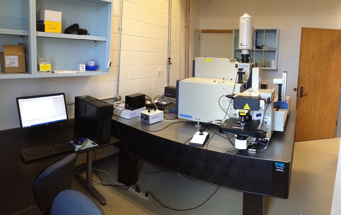
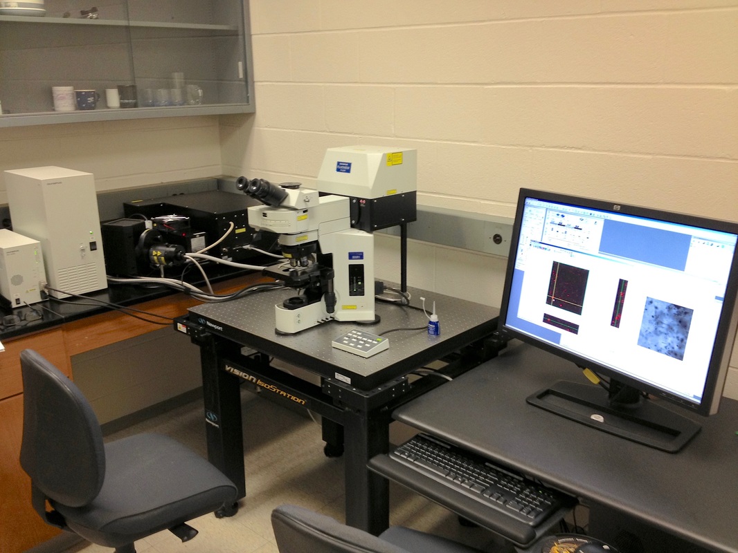
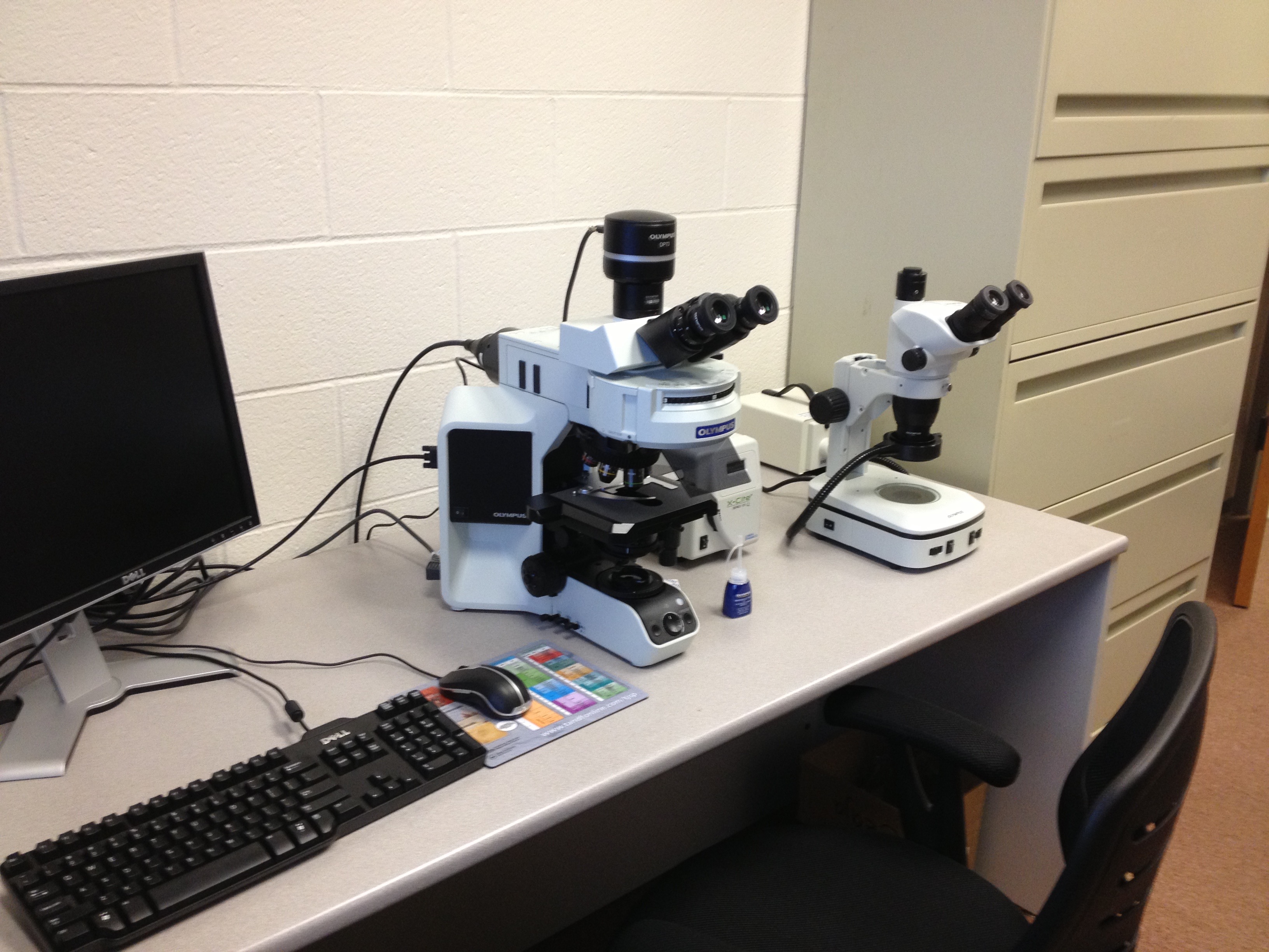
These are the instruments in my lab I use to study fossil microorganisms and the carbonaceous material and minerals of which they are made. Raman spectroscopy provides in situ, micron-scale chemical information about the organic remains of the microbes, as well chemical information of the host matrix. Combined, the Raman spectra and the 2-D and 3-D optical/chemical images produced by these techniques provide greater amounts of morphological and biochemical information than could be attained by other, more traditional means. Most important for very ancient fossil structures, these techniques can provide powerful evidence that the remains are in fact those of microorganisms.
|
Raman spectrometry
Organic molecular structural analyses and mineral identification
|
|
Confocal Laser Scanning Microscopy
2-D and 3-D fluorescence imaging of organic fossils
|
|
Light Microscopy
Optical and UV fluorescence imaging of fossils and minerals
|
